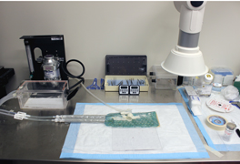
The laboratory includes a full setup for in vivo imaging and intravital microscopy capable of imaging length scales from macroscopic (25 cm x 25 cm) down to subcellular resolution. A stainless steel preparation table containing a circulating water warming pad and isoflurane vaporizer is centered between a Licor Odyssey CLx near-infrared imager for macroscopic, organ, and tissue slice imaging and an Olympus FV 1200 upright confocal microscope for intravital microscopy.
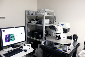
The Olympus FV 1200 Confocal Microscope contains both a solid state 635 nm and a 748 nm laser for imaging probes in two separate near-infrared channels. In addition, the microscope contains a solid state 405 nm laser, an Argon laser with 458 nm, 488 nm, and 514 nm laser lines, and a 542 nm HeNe laser. These visible light channels can be used to image fluorescent proteins (e.g. H2B-RFP to label the nuclei of tumor cells), vascular probes, and additional injectable agents such as fluorescent antibodies providing context to agent distribution. The system is mounted on an anti-vibration air float table. A motorized stage allows point visiting and time-lapse imaging during and after injection of fluorescent agents. 5X, 20X, and 60X objectives enable fields of view from beds of vascularized tissue down to single cell and subcellular distribution of biologics, nanoparticles, and other fluorescent agents.
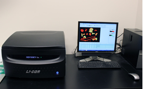
The Licor Odyssey CLx imager contains dual wavelength excitation at 685 or 785 nm for near-infrared imaging. The scanner resolution and imaging field is adjustable down to 21 microns per pixel, overlapping with the capabilities of the confocal microscope. The detectors have a dynamic range over 4 orders of magnitude allowing quantitative detection of fluorescence from thick tissues such as ex vivo organs, tissue slices (0.5 mm sections), and histology slices (~ 5 microns).
Other equipment:
Additional equipment in the lab includes a Shimadzu Prominence semi-preparative HPLC with analytical and preparative C18 and size exclusion columns, a refrigerated Sorvall Legend X1R centrifuge, Eppendorf microcentrifuge, rotary evaporator, molecular biology capabilities (protein and DNA gel boxes, dry transfer system for Western blotting, shaker, incubator, etc.), and 2 chemical hoods.

The Thurber Lab is located in the University of Michigan North Campus Research Complex, home to several core facilities.